
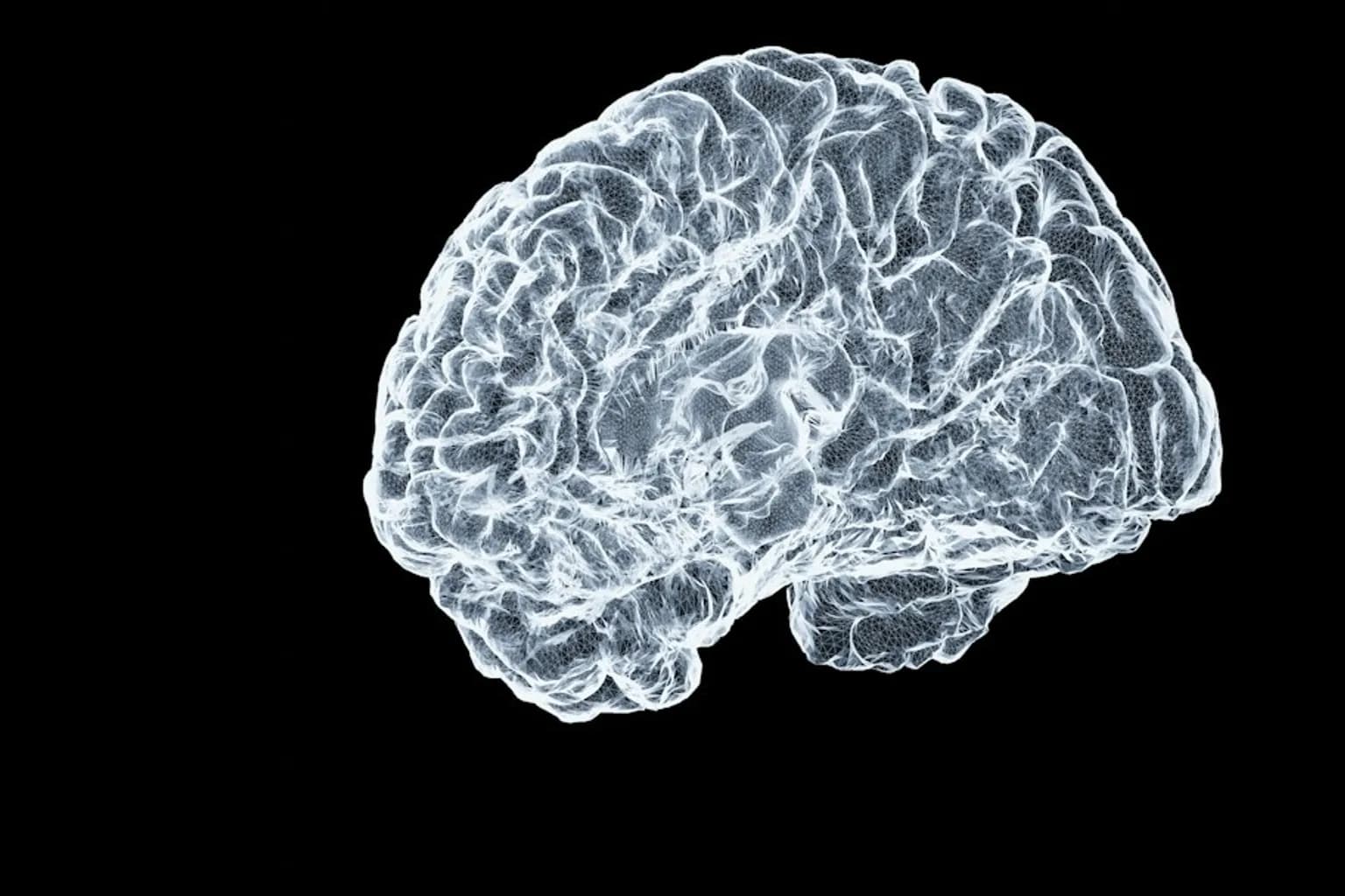
Scientists at Stanford Medicine have found that the human brain develops through two distinct cellular systems, challenging the long-standing model in which a single early progenitor cell gives rise to the whole organ. The findings, published in Nature Neuroscience on September 18, 2026, trace the brain's front and back regions to separate cell populations that follow genetically locked developmental paths from the earliest embryonic stages.
One population expresses a gene designated Otx2. It is exclusively committed to forming the forebrain and midbrain. The other expresses Gbx2 and develops solely into the hindbrain. The two populations never overlap, according to the researchers, including at the earliest points in development.
The distinction also extends to the way their DNA is packaged. The team found that the two progenitor populations have entirely different DNA configurations, indicating that their developmental programmes are separate from the outset.
The Otx2-linked system gives rise to regions associated with language, mathematics, abstract thought and reflection on existence. The midbrain and forebrain also govern consciousness, logic, vision, hearing and movement control. The Gbx2-linked system produces the hindbrain, which sits at the base of the skull and manages survival functions including heart rate, breathing and sleep cycles, as well as swallowing and digestion.
Kyle Loh, PhD, an associate professor of developmental biology and the research's senior author, and his colleagues identified the two fundamentally different forms of brain progenitor cell while studying mouse embryos during gastrulation. Their work indicates that the front of the brain arises from a wholly different progenitor cell from the back.
The researchers found the same developmental pattern in chickens, zebrafish and acorn worms. They said the arrangement of two separate brain organs arose at least 550 million years ago and has been retained across species. The study establishes the pattern in humans, mice, chickens, zebrafish and acorn worms, though it remains unclear whether it is present in all vertebrate lineages.
The finding has a practical consequence for laboratory research. Scientists have struggled for decades to grow hindbrain neurons because they began with the wrong progenitor cells, the Stanford team said. By following the Gbx2 developmental route, the researchers successfully transformed human pluripotent stem cells into functional hindbrain motor neurons for the first time.
These laboratory-grown neurons produced electrical activity and proteins associated with control of facial and swallowing muscles. The method gives researchers a way to study cells affected in ALS, spinal muscular atrophy and other brainstem diseases, and may support the testing of treatments directed at those conditions.
The work leaves key questions unresolved. Researchers do not yet know how the two systems functionally integrate, including whether specific anatomical pathways link them. They also do not know why evolution maintained the dual architecture when a single unified system might appear more efficient, or how many diseases beyond ALS and spinal muscular atrophy can be examined through the new culture method.




Comments (0)