
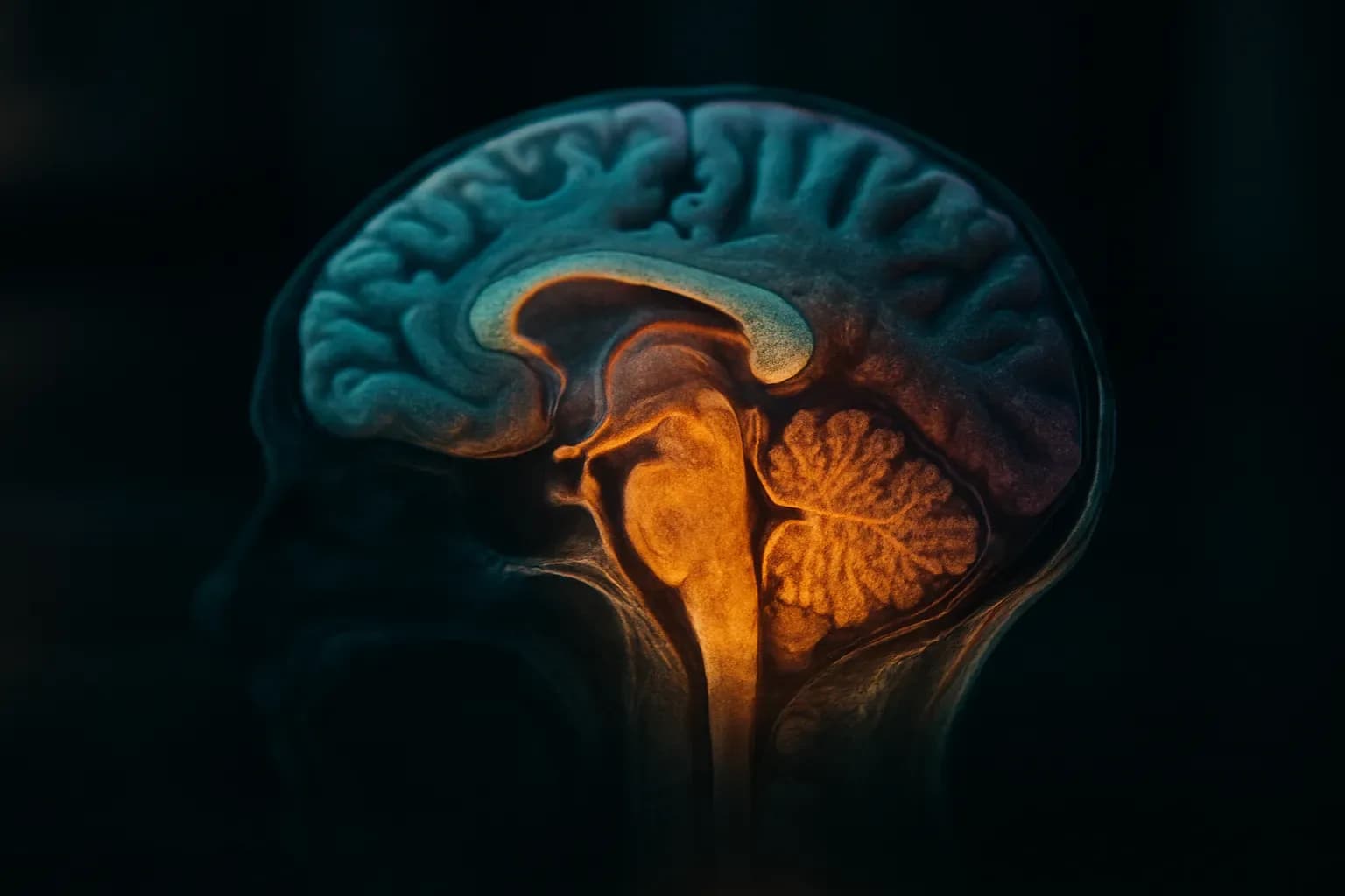
At EPFL, Giovanni D'Angelo led a team that examined thin slices of mouse brain tissue point by point with mass spectrometry imaging, at micrometric resolution. D'Angelo is a professor at The Kristian Gerhard Jebsen Foundation Chair in Nutrition and Metabolism at EPFL, while Luca Fusar Bassini and Gioele La Manno collaborated on the project.
The researchers measured 172 different lipids in 109 brain slices from 11 mice, producing approximately 7 million measurements.
They then used artificial intelligence and machine learning models to turn the two-dimensional scans into a three-dimensional atlas. The analysis identified 539 chemically distinct regions, termed “lipizones”, each defined by its own mix of lipids.
The result is the Lipid Brain Atlas, described as the first detailed 3D map of lipids in the brain. Its patterns were distinctive enough for the researchers to identify where a tissue sample came from by examining its lipids alone.
The most striking finding concerns white matter. Often treated as relatively uniform connective tissue, it emerged as a patchwork of separate chemical territories.
Some of those territories align with established brain structures and cell types. Others connect brain regions that are far apart, including lipizones linking clusters of nerve cell bodies with areas reached by their fibres. Gene-based maps have usually missed those links.
“In effect, lipids work like postal codes, giving distant but related parts of the brain the same chemical address,” D'Angelo said.
The atlas also identified chemical borders in the choroid plexus, the tissue that makes the brain's fluid, and in the linings of the brain's internal cavities.
Lipids form neuron membranes, provide myelin around nerve fibres and help brain cells send and receive signals. They make up the majority of the dry weight of the human brain.
Fusar Bassini said the findings changed the picture of the cells that make myelin. “This shows that the cells which make myelin are far more varied in their chemistry than their genes alone would suggest.”
The team also looked at pregnancy in mice. It found substantial shifts in lipid composition across numerous brain regions, particularly in the cortex, while levels of galactosyl ceramide rose sharply through much of the brain's white matter.
“The Atlas reveals that the brain constantly adjusts its lipids to meet the body's changing needs,” La Manno said.
Changes in brain lipids are increasingly linked to conditions ranging from depression to Alzheimer's disease. The findings were published in Nature, and the Lipid Brain Atlas is freely available online.





Comments (0)